2026.03.05 Thursday
Real-Time Imaging of Microplastics in the Body Improves Understanding of Health Risks
Scientists develop fluorescent dye-loaded nanosized, irregularly-shaped microplastics to track their movement in real time, following ingestion, through deep-tissue imaging
Microplastics (MPs), defined as plastic fragments with sizes ranging from millimeters (<5 mm) to nanometers, have become a growing environmental and public health concern. First identified in the 1970s, these particles are now omnipresent in water, soil, air, and everyday products, such as detergents and cosmetics. Hundreds of these particles can be ingested or inhaled in a day, with smaller particles posing a greater risk as they may accumulate in organs such as the liver, lungs, kidneys, and even the brain. Understanding the in vivo behavior and biological effects of these irregularly shaped nano-sized MPs is therefore critical.
To this end, researchers led by Associate Professor Masakazu Umezawa from the Department of Medical and Robotic Engineering Design, Tokyo University of Science, Japan, have developed fluorescent MPs that emit light in the second near-infrared (NIR-II) biological window, enabling real-time deep-tissue imaging. The research team included a second-year master's student Mr. Ryo Nagasawa, a recent Master's graduate Mr. Sota Inoue, and Professor Kohei Soga, also from the Department of Medical and Robotic Engineering Design, Tokyo University of Science, Japan. Their work was published online in the journal Environmental Science: Advances on February 10, 2026, as part of the HOT Articles in Environmental Science: Advances collection.
"The issue of MPs has been raised worldwide, and there are several news articles on the web, but the topic of how they move inside the body has not been discussed, and there remain many unclear aspects. I wanted to contribute by proposing a new method to clarify this issue," says Assoc. Prof. Umezawa.
When examining the impact of MPs in vivo, a key factor is their shape. In the real world, plastics are weathered and crushed into irregularly shaped fragments, which may behave differently inside the body than the smooth, artificial spherical particles used as model MP samples in most studies. To create more realistic models, the researchers had developed a method to synthesize irregularly shaped, fluorescent poly(ethylene terephthalate) (PET) nano-MPs and demonstrated their feasibility for real-time tracking in mice. This work was made available online on December 23, 2025 and was published in Volume 28, Issue 1 of the Journal of Nanoparticle Research on January 1, 2026. They have now further improved this synthesis method to include other common plastics: polypropylene (PP), polyethylene (PE), and polystyrene (PS).
This method involved fragmenting plastic granules in a solvent into nanosized particles, which were then mixed with the fluorescent dye, IR-1061. For PET, the dye diffused readily as the solvent caused the plastic to swell. However, PP, PE, and PS were less compatible, so the mixture was gently heated to 55 °C, causing the polymer chains to expand and allowing the dye to enter. Adding bovine serum albumin prevented clumping and shaped the particles into an irregular form, resulting in water-dispersible particles between 30 and 300 nanometers in size. These particles retained more than 80% of their fluorescence for at least 30 days, making them well-suited for long-term tracking studies.
Fluorescent imaging following oral administration in mice showed that these MPs remained in the stomach for several hours before migrating to the intestines and were subsequently excreted in feces. No fluorescence was detected in tissues outside the gastrointestinal tract, indicating negligible intestinal absorption. Notably, particles' sizes influenced their intestinal retention, with the smallest particles showing longer retention times.
The team further demonstrated the platform's versatility by successfully loading the irregularly shaped MPs of PET, PP, and PE with another fluorescent dye, Nile red, to study their cellular uptake in vitro. In mouse fibroblasts, the MPs were taken up at concentrations as low as 2.0 µg/mL, a fraction of the amount reported for spherical particles. These findings have been published in the journal Environmental Science: Advances on February 18, 2026.
With global plastic waste projected to rise from 188 million tons in 2016 to 380 million tons by 2040, understanding the fate of MPs in the body is more urgent than ever. This method of creating fluorescent-loaded MP models that resemble real-world plastics in morphology enables the study of chronic exposure effects, the different routes of entry, and their interaction with biological tissues over time.
"The development of methods for synthesizing NIR-II-fluorophore-loaded microplastic models with various chemical compositions will support risk assessments by providing insights into the environmental and biological fate of MPs," says Assoc. Prof. Umezawa.
These findings lay the foundation for further research on how this method may help regulators better evaluate health risks due to microplastics in food and air.
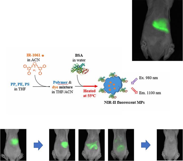
Image title: Real-time tracking of microplastics in mice using second near-infrared (NIR-II) fluorescence imaging
Image caption: Microplastic particles of polypropylene, polyethylene, and polystyrene were loaded with IR-1061 and orally administered to mice. NIR-II imaging under 980 nm irradiation tracked their gastrointestinal accumulation and subsequent excretion from 30 minutes up to 48 hours.
Image credit: Dr. Masakazu Umezawa from Tokyo University of Science, Japan
License type: Original content
Usage restrictions: Credit must be given to the creator. Cannot be reused without permission.
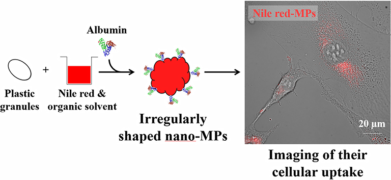
Image title: Nile red-loaded microplastics and their cellular uptake
Image caption: Nano-microplastic particles loaded with Nile red dye enabled high-sensitivity imaging of cellular uptake. In experiments with mouse fibroblasts, the dye-loaded particles achieved clear visualization at concentrations as low as 2.0 µg/mL.
Image credit: Dr. Masakazu Umezawa from Tokyo University of Science, Japan
Image source link: https://pubs.rsc.org/en/content/articlehtml/2026/va/d6va00031b
License type: CC BY-NC
Usage restrictions: Credit must be given to the creator. Only non-commercial uses of the work are permitted.
References
| Title of original paper | : | Synthesis of near-infrared-fluorophore-loaded microplastics with different compositions for in vivo tracking |
| Journal | : | Environmental Science: Advances |
| DOI | : | 10.1039/D5VA00360A |
About The Tokyo University of Science
Tokyo University of Science (TUS) is a well-known and respected university, and the largest science-specialized private research university in Japan, with four campuses in central Tokyo and its suburbs and in Hokkaido. Established in 1881, the university has continually contributed to Japan's development in science through inculcating the love for science in researchers, technicians, and educators.
With a mission of "Creating science and technology for the harmonious development of nature, human beings, and society," TUS has undertaken a wide range of research from basic to applied science. TUS has embraced a multidisciplinary approach to research and undertaken intensive study in some of today's most vital fields. TUS is a meritocracy where the best in science is recognized and nurtured. It is the only private university in Japan that has produced a Nobel Prize winner and the only private university in Asia to produce Nobel Prize winners within the natural sciences field.
■
Tokyo University of Science(About TUS)
![]()
About Professor Kohei Soga
from Tokyo University of Science
About Associate Professor Masakazu Umezawa
from Tokyo University of Science
Dr. Masakazu Umezawa serves as an Associate Professor in the Department of Medical and Robotic Engineering Design, Faculty of Advanced Engineering, Tokyo University of Science, Japan. Over the years, he has published 169 papers in high-impact factor scientific journals. His main research interests include nanomaterials chemistry, protein-nanomaterial interactions, and environmental physiology. He has won several awards and honors, such as the "Most Cited Paper Award" and "Hot Article Award 2022" in recent years. He has been associated with multiple academic societies, including the Japan Society of Bioimaging and the Japanese Biochemical Society.
Funding information
'Synthesis of near-infrared-fluorophore-loaded microplastics with different compositions for in vivo tracking' and 'Near-infrared (NIR-II) fluorescent poly(ethylene terephthalate) nano-microplastics for in vivo tracking': The work was in part supported by the Japan Society for the Promotion of Science (JSPS) KAKENHI (Grant numbers: 22K06565, 23K24593, and 25K02871).
'Preparation of Irregularly Shaped, Nano-Sized, Fluorescent Microplastic Particles for Tracing Cellular Uptake': This work was in part supported by JSPS KAKENHI (Grant numbers: 22H03335, 22K06565, and 25K02871).